I am neither especially clever nor especially gifted. I am only very, very curious.
I have much interest in research. Since second year in university, I have entered several labs and have done quite a few projects and research work.
My undergraduate research interest includes image processing, computer aided diagnosis, Traditional Chinese Medicine, complex network, electronics, data mining.
Now I have joined Department of Electrical and Computer Engineering, University of Washington, Seattle to continue my research on medical image processing. I am currently working on developing image processing and artificial intelligence techniques to assist vascular image review and clinical diagnosis. Introduction to my research in 10 slides ![]() Research in 10 slides.pdf, 25 slides:
Research in 10 slides.pdf, 25 slides: ![]() Research in 25 slides.pdf
Research in 25 slides.pdf
I would consider myself as a medical researcher as well as a technical innovator, so it would be easier to understand my contributions to both the medical and technical societies from two outlines.
Contributions to medical science
I am interested in researching on human vasculature and vascular diseases. Sufficient blood supply to the brain is important for maintaining health, and vascular diseases are among top death causes worldwide (such as stroke), but the current gap is we are lacking sufficient techniques to quantitatively and automatically explore human vasculature. MRI is an ideal modality to have a better view of arteries (even wall) but at great cost of manual reading. By introducing image processing and machine learning techniques, I made many labour intensive projects feasible and led to new discoveries in medical science.
1. iCafe for intracranial artery quantification and analysis: iCafe page (est reading time: 5 min)
Validated the feasibility of artery quantification from MRA in the MRI paper.
Developed iCafe to quantify intracranial arteries in the MRM paper.
Found vascular reduction through aging among elderly population in the NoA paper.
Identified vascular changes before and after carotid revascularization: ![]() ISMRM2019_Arizona.pdf
ISMRM2019_Arizona.pdf
Validated the strong correlation of vascular features from MRA with blood flow in another MRI paper
Similar approach to quantify and analyze peripheral arteries: ![]() ISMRM2019-pCafe.pdf
ISMRM2019-pCafe.pdf
Explored the arterial collateralization differences on peripheral artery disease: ![]() ISMRM2019_PAD.pdf
ISMRM2019_PAD.pdf
Discovered reduced intracranial arteries might be an indicator for dementia: ![]() ISMRM2019_ACT.pdf
ISMRM2019_ACT.pdf
2. FRAPPE for popliteal vessel wall quantification and analysis: FRAPPE page (est reading time: 5 min)
Validated the feasibility of fully automated popliteal vessel wall analysis from MR knee scan in the MRM paper.
Segmented vessel wall on 3.5 million popliteal images from the OAI dataset and discovered vessel wall remodeling patterns. (JAHA paper)
Discovered the correlation between physical exercises and popliteal lumen diameters. (under review)
Innovations in technical developments
I have great interest in extracting and understanding the information from medical images. Image processing and machine learning (especially deep learning) techniques are perfect tools to help my explorations. I am able to apply novel techniques to solve clinical problems, and keep improving the performance of techniques during applications. My technical development is to fill the technical gap out of clinical needs instead of chasing higher evaluation metrics.
1. Techniques to improve the performance of vessel segmentation
Y-net, one of the earliest deep learning approaches for intracranial artery segmentation. BIBM paper
Fully automated vessel wall analysis by 3D artery localization (tracklet refinement after artery detections) and vessel wall segmentation in the polar coordinate system. IEEE paper
Designed confidence scores from neural network predictions for segmentations. CIRCGEN paper
2. Techniques for machine learning based vascular disease diagnosis/screening
Automated image quality assessment from weighted patches. SPIE paper
Carotid lesion classification using domain adaptive deep learning techniques. ISMRM abstract, MRM paper
Visualizing and understanding feature map of popliteal plaques. ![]() ISMRM2020_plaque.pdf
ISMRM2020_plaque.pdf
Stenosis detection from CT images in multiplanar view. ![]() ISMRM2020_stenosis.pdf
ISMRM2020_stenosis.pdf
3. Techniques for automated intracranial arteries analysis
Simultaneous artery tracing and segmentation by cross-sectional plane matching. MICCAIW paper
Deep open active contour model for artery tracing. MICCAI paper
Artery refinement on MRA with heavy motions and artifacts (infants). JNM paper
Graph neural network labeling for intracranial arteries. MICCAI paper
Personal statements about my research style
There is a huge gap between medical and technical societies (both ideologically and practically), my aim is to bridge between technical and medical world.
What to do is always more important than how to do it. Good idea + Data + AI lead to impactful medical imaging research.
Technical development solving an unmet challenge is far more meaningful than patching on an existing method to solve the same problem. I hate those boring derivatives of U-Net without a clear aim.
AI is not a black box, and we are still at the beginning stage of understanding AI. End to end training might work well, but I'd rather break the challening task into steps and understand how AI behaves in each step.
A good machine learning engineer can not only tune the model to increase its performance, but also understand the underlying mechanism of the task, and use human knowledge to guide model design and training.
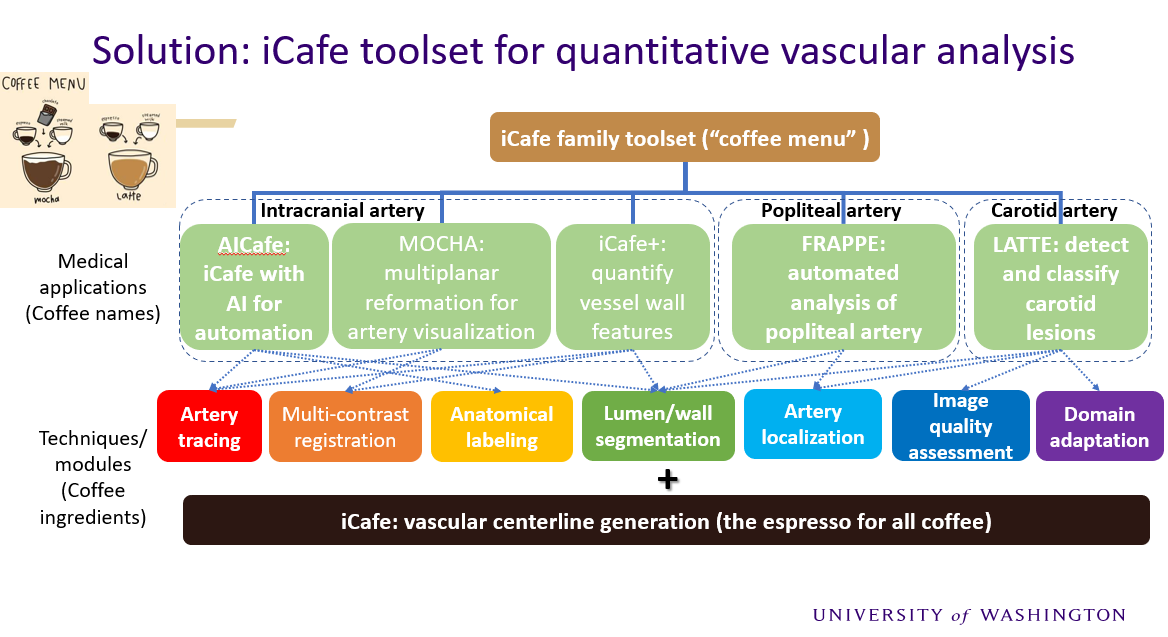
The tools I developed for vascular analysis are all related with coffee names starting with iCafe. Here is a chart showing main modules in the iCafe family toolset.

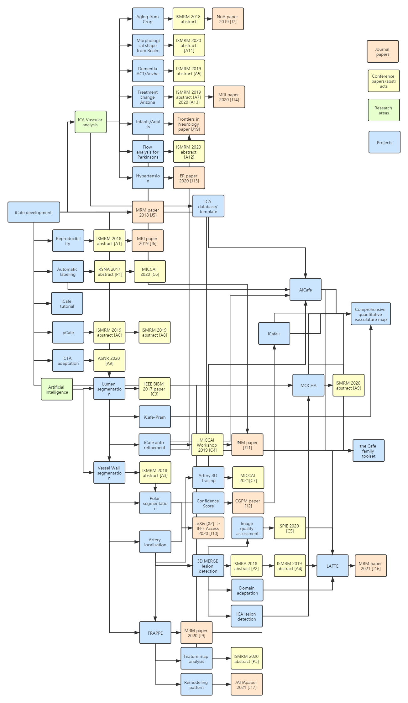
The following chart is for relations of projects and publications during my graduate school research.

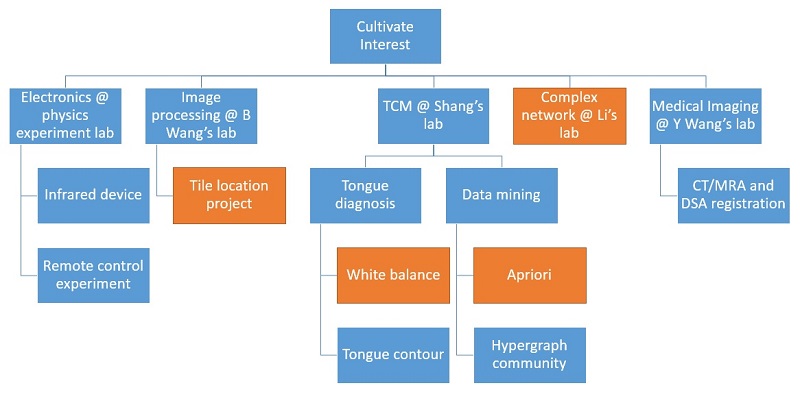
The following chart is for my undergraduate research.

Main research experience
2013.9 ~ 2014.12 Research in Fudan Physics Teaching Lab with Professor and Lab director Yongkang Le, mainly about application of remote control lab applications, chip based electronic devices.
2013.10 ~ 2016.6 Research with Associate professor Huiliang Shang on projects about image processing, medical topics on Traditional Chinese medicine, robotic control, visible light positioning, circuit theory.
2013.10 ~ 2014.11 Guided by professor Bin Wang through The FDUROP project about image processing , an undergraduate research funding project on image processing, computer vision and pattern recognition.
2014.3~2015.1 Research on immunization strategy with Professor Xiang Li, in Adaptive Networks and Control Lab.
2015.8 ~ 2016.6 Research with Professor Yuanyuan Wang and Yi Guo, doing my capstone project about medical image registration, a research using network structure and circuit simulation methods to perform image registration.
2016.6 ~ 2016.7 Research in Center for Biomedical Imaging Research, Tsinghua University with Huijun Chen. Quantitative analysis of intracranial arteries through aging.
2020.6 ~ 2020.9 Research intern in United Imaging Intelligence America (Boston) with Dr. Shanhui Sun and Terrence Chen. Landmark tracking for stent enhancement.
2021.5 ~ 2021.8 Research intern in Genentech (South San Francisco) with Dr. Reza Negahdar for imaging and non-imaging feature fusion.
2016.9 ~ 2021.8 Co-advised by Professor Chun Yuan and Jenq-Neng Hwang in the UW ECE PhD program.
2021.10 ~ Now Research scientist at Philips Research North America. Lung ultrasound (Lumify) AI analysis.
Please see my Curriculum Vitae for more information.
Click subtopic links above to see articles about the progress and outcome of my research.
Email me if you have interest in detailed information about the paper.
Updated on Dec. 2022