I gave an invited talk at Imaging Elevated symposium this week about FRAPPE.
I think the take home message is not just how impressive AI is at drawing contours and saving manual labors. We should think more about how AI can transform our medical imaging research from this project.
As a quick reminder, the beauty of this popliteal vessel wall analysis project is in three dimensions. First, popliteal artery is a vascular bed which few people have paid attention to but actually very informative (atherosclerosis is a systemic disease). Luckily, the popliteal MR data sit there. Second, the vessel wall analysis is not possible without help from AI. It will take 63 years for an expert to finish the annotations. Third, AI is able to handle this task with limited amount of training labels and perform equally well compared with human.
People doing medical research need clever minds to propose interesting hypothesis to enhance our knowledge about unknown mechanism about human body and diseases. Identifying a meaningful, innovative and urgently needed research topic is important, not only for earning funding to support the research, but also adding values to your life (nobody wants to waste their life on nonsense or repeating other people’s work).
One unique challenge for medical research is that it has so many uncertainties and variables. Thus, a large enough dataset is the ideal way to test our assumptions and make solid conclusions. That is why big data is usually associated with prestigious publications. Enough data points ensure significance for statistical analysis, and supports your hypothesis. Also comprehensive features allow a complete analysis of data.
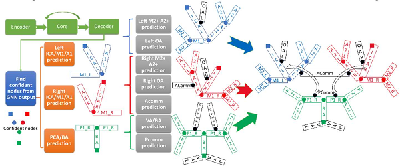
However, for imaging studies, the information is multi-dimensional. Just as the saying goes "A picture is worth a thousand words", when we have a large image dataset, the features contained in the image dataset are not easily quantifiable for easy modeling or statistical analysis. Luckily, finding signal from large dimensional data is actually where deep learning models are good at. Through layer of convolutions, useful features are kept while noises are sifted out, so finally we can build powerful models for classification, regression, segmentation, etc. That is also the reason why medical imaging / radiology area soon became crazy about AI in recent conferences. Recently, better understanding of deep learning behaviors facilitate stronger models which uses few annotations while having good performances. More importantly, advances in AI techniques make fully automated process on medical images possible. That brings to the topic of why FRAPPE is unique. “FR” represents fully automated and robust. Compared with previous vessel wall analysis tools with AI component, FRAPPE is fully equipped with AI for automation. For example, previous methods usually take it for granted that manual preprocessing with region of interest identification before vessel wall analysis is a given condition, but if a human needs to identify the location of 3.5 million popliteal images, even the preprocessing takes as little as 1 second, it will drive any human crazy. So we are not only expecting AI to process large number of images, we also want it to be fully-automated.
I believe most of the medical researchers have a brilliant brain to figure out what is the most critical gap in current understanding to medicine. And if you are lucky, you might find data for your research by either having enough funding for large population study or having access to an existing dataset. What is left is (finding a hard working Phd student for) developing fully automated AI techniques on image analysis.
admin1
memeda
ssy
 Login
Login