Research
Research Overview
Building clinically useful medical AI through data-efficient learning, knowledge-guided modeling, and quantitative imaging.
I am a Research Scientist in Philips Research / Philips Ultrasound in Cambridge, Massachusetts. My work applies machine learning and computer vision to medical imaging, with an emphasis on translating algorithms into tools that can support clinical interpretation and research. My current focus is AI for point-of-care lung ultrasound; my doctoral research at the University of Washington centered on quantitative vascular image analysis from MRI. My CV and full publication list provide the complete career and publication record.
1. Clinical translation: AI for point-of-care lung ultrasound
At Philips, I contribute to AI-assisted analysis of lung ultrasound on the Lumify point-of-care ultrasound platform, targeting findings including lung consolidation and pleural effusion. The work spans data-efficient model development, interpretation of ultrasound video, aggregation of findings across anatomical lung zones, and clinical evaluation. My CV summarizes my role in the development cycle and clinical-translation milestones.
Learning with limited annotations
Ultrasound data can be expensive to annotate at frame level, and labels may be available at different granularities. The IPMI paper Weakly Semi-Supervised Detection in Lung Ultrasound Videos studies learning from frame-level labels, video-level labels, and unlabeled data. In my CV's Philips research summary, I report an estimated reduction in annotation effort for 15,000 videos from 15,000 hours to approximately 250 hours using a teacher–student mutual-learning framework.
To exploit unlabeled data, the ISBI paper Contrastive Self-Supervised Learning for Spatio-Temporal Analysis of Lung Ultrasound Videos investigates contrastive pretraining for ultrasound video representations. The reported experiment used 27,063 unlabeled videos from 528 patients and improved test AUC from 0.65 to 0.88 when only 5% of the labeled data was used; the dataset and result are documented in the publication and CV.
At the detection stage, the paper Graph-Based Pseudo-Label Selection for Weakly Semi-Supervised Object Detection in Ultrasound Videos uses graph structure to help select pseudo-labels when box annotations are limited.
Aggregating evidence from frames to videos and patients
A single ultrasound frame rarely captures all the evidence needed for a reliable decision. My graph-based work explores how to combine detections across time and anatomy rather than relying only on isolated frame predictions or simple pooling.
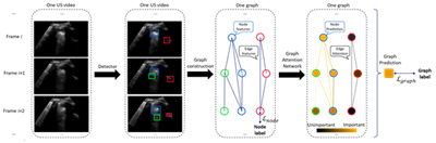
GADA — Graph Attention-based Detection Aggregation for Ultrasound Video Classification. GADA represents detected regions as nodes in a spatiotemporal graph and uses graph attention to aggregate local predictions into video-level classification. See the project/paper page, the ICCV Workshop paper, and its arXiv preprint. The CV summary reports AUCs of 0.943 for consolidation and 0.960 for pleural effusion across six sites and more than 9,900 lung ultrasound videos.
ZAGNet — Zone-Aware Graph Aggregation Network for Patient-Level Lung Ultrasound Diagnosis. ZAGNet represents temporally tracked findings as graph nodes and uses anatomical adjacency between lung zones to support patient-level classification, including examinations with missing zones. The project/paper page includes the method and results, and the conference PDF provides the paper. On a multicenter dataset of 714 subjects and 20,256 video loops, the reported AUCs were 0.803 for consolidation and 0.893 for pleural effusion, compared with max-pooling AUCs of 0.677 and 0.804 (mean-pooling AUCs were 0.674 and 0.815) (paper page).
Clinical usefulness also needs to be evaluated with readers, not only with model-level metrics. The clinical-evaluation summary in my CV describes a multi-reader, multi-case study in which AI assistance met non-inferiority and superiority endpoints, increased reader-averaged AUC by 0.101 for consolidation and 0.046 for pleural effusion, and reduced reading time by 16.6% (12.5 seconds).
Together, these projects form a progression from data-efficient detection to video-level reasoning and patient-level aggregation, with the supporting papers and project pages linked above.
2. Knowledge-guided AI for challenging medical images
A recurring technical theme in my research is to turn domain knowledge into computational structure. Depending on the problem, that structure may encode anatomical hierarchy, geometric shape, tracking continuity, or relationships across time and space.
Anatomical labeling of intracranial arteries. The MICCAI paper Automated Intracranial Artery Labeling using a Graph Neural Network and Hierarchical Refinement models hierarchical relationships among artery segments to make anatomical labeling more consistent with the vascular tree.
Vessel tracing with shape constraints. The MICCAI paper Deep Open Snake Tracker for Vessel Tracing develops a deep-learning-based active-contour approach for tracing vessels. Related work on simultaneous artery tracing and segmentation uses joint optimization across multiplanar reformations; see the detailed paper page.
Vessel-wall localization and segmentation. The paper Automated Artery Localization and Vessel Wall Segmentation of Magnetic Resonance Vessel Wall Images using Tracklet Refinement and Polar Conversion combines tracklet refinement for 3D localization with a polar-coordinate representation that exploits the approximately circular cross-section of vessels.
Spatiotemporal and anatomical reasoning in ultrasound. The graph structures in GADA and ZAGNet extend this same principle from vessel anatomy to temporally connected detections and relationships among lung zones.
The common idea is not to add complexity for its own sake, but to encode task-relevant structure that may improve data efficiency, robustness, and the interpretability of model outputs. The papers above provide concrete method descriptions and experiments; the full publication list collects the broader record.
3. Quantitative vascular imaging for scientific discovery
During my Ph.D. in Electrical and Computer Engineering at the University of Washington, I developed image-analysis tools that convert vascular MRI into structured quantitative measurements. These projects aimed to make analyses reproducible at scale and to support clinical research beyond manual image review.
iCafe — intracranial artery feature extraction. iCafe is a software platform for tracing, labeling, visualizing, and quantifying the cerebral arterial network from MRA. The project page explains its workflow and feature extraction; the foundational paper on quantitative intracranial vascular feature extraction and the older-adult vascular assessment study describe validation and application. My CV records use of iCafe by more than 34 sites across 9 countries.
FRAPPE — popliteal artery and vessel-wall analysis. FRAPPE is an automated pipeline for locating popliteal arteries and measuring vessel-wall features from routine knee MRI. The project overview and technical paper page describe the methodology. The work was applied to approximately 3.5 million popliteal artery images from the Osteoarthritis Initiative; see also the peer-reviewed paper.
LATTE — AI-assisted carotid atherosclerotic lesion screening. LATTE combines automated analysis of 3D carotid MRI with domain-adaptation methods to support screening for atherosclerotic lesions. The project overview describes the workflow, and the peer-reviewed Magnetic Resonance in Medicine paper reports the associated technical evaluation.
These tools illustrate how computer vision can be used not only to automate individual tasks, but also to generate quantitative imaging features for studies of aging, cognition, and vascular risk. The iCafe application papers, FRAPPE paper, and LATTE paper provide examples of this translational-to-scientific pathway.
4. Earlier work in medical image analysis
My earlier projects explored related problems in different settings. The undergraduate capstone project on medical image registration using network structure and circuit simulation represented vascular trees as graphs and used structural features to guide image matching. I also worked on traditional Chinese medicine image analysis, including the paper on automatic tongue-image segmentation using color enhancement in L*a*b* space. These projects are part of the longer research trajectory documented in my CV and publication list.
Building clinically useful medical AI by combining computer vision, data-efficient learning, and medical knowledge.
I am a Research Scientist in Philips Research / Philips Ultrasound in Cambridge, Massachusetts. With more than 13 years of research experience across academia and industry, I develop machine-learning and computer-vision methods that turn complex medical images into reliable, clinically useful information. My work spans point-of-care lung ultrasound and vascular MRI. Across these areas, I focus on the practical challenges that determine whether medical AI can be useful beyond a benchmark: limited annotations, domain shift, anatomical and spatiotemporal structure, inter-reader variability, and clinical validation.
I received my Ph.D. in Electrical and Computer Engineering from the University of Washington, where I worked in vascular imaging with Professors Chun Yuan and Jenq-Neng Hwang.
1. Translational AI for clinically useful medical solutions
At Philips, I contribute to the development of AI-assisted pulmonary-abnormality analysis for the Lumify ultrasound platform, including lung consolidation and pleural effusion. My work spans data-efficient learning and clinical translation: weakly and semi-supervised learning to reduce dependence on frame-level labels; contrastive self-supervised learning to learn from unlabeled videos; and detection, tracking, and graph-based models that aggregate local findings into video- and patient-level evidence.
A central challenge is that a diagnosis rarely depends on a single frame. Reliable analysis must combine noisy detections over time and across anatomical lung zones, while accounting for incomplete scanning protocols and limited labeled data.
Selected examples
Data-efficient learning. Contrastive pretraining on 27,063 unlabeled ultrasound videos from 528 patients improved test AUC from 0.65 to 0.88 when using 5% of the labeled data.
Reduced annotation burden. A teacher–student mutual-learning framework was designed to use frame-labeled, video-labeled, and unlabeled data; the estimated annotation effort for 15,000 videos was reduced from 15,000 hours to approximately 250 hours.
Clinical evaluation. In a multi-reader, multi-case study, AI assistance improved reader-averaged AUC by 0.101 for consolidation and 0.046 for pleural effusion, while reducing interpretation time by 16.6%.
Structured evidence aggregation. GADA models spatiotemporal relationships among detections for video-level classification, while ZAGNet uses anatomical relationships among lung zones to support patient-level diagnosis.
The goal is not simply to classify images, but to develop systems that can operate with realistic data and workflow constraints and be evaluated for their value to clinicians.
2. Knowledge-guided AI for challenging medical images
I aim to encode relevant medical knowledge into model representations instead of treating images as generic arrays. Depending on the problem, this can mean representing anatomical relationships as a graph, enforcing geometric constraints, using shape-aware coordinates, or tracking structures across slices and time.
Anatomical artery labeling: graph neural networks and hierarchical refinement to identify intracranial artery segments in a way that reflects vascular anatomy.
Artery tracing: optimization and active-contour constraints to improve tracing and segmentation of vessels in three-dimensional MRA.
Vessel-wall segmentation: polar-coordinate representations that exploit the approximately circular geometry of vessels.
Robust localization: tracklet refinement to connect detections across slices into coherent three-dimensional artery paths.
This knowledge-guided approach continues in lung ultrasound, where graph-based methods represent temporal continuity and anatomical relationships among findings. I use structure not as decoration, but as a way to make learning more data-efficient, robust, and clinically meaningful.
3. Quantitative vascular imaging for scientific discovery
During my Ph.D. at the University of Washington, I developed automated and semi-automated tools for extracting quantitative vascular features from MRI. These projects illustrate a broader aim of my research: using image analysis not only to automate review, but also to make large-scale clinical investigation possible.
iCafe — intracranial artery feature extraction. A software platform for tracing, labeling, visualizing, and quantifying the cerebral arterial network from MRA. It has supported studies of vascular morphology in relation to aging, cognitive impairment, and vascular risk, and has been used by more than 34 sites in 9 countries.
FRAPPE — popliteal artery vessel-wall analysis. A fully automated pipeline for locating popliteal arteries, segmenting vessel walls, and measuring vascular features in routine knee MRI. It enabled analysis of approximately 3.5 million images from the Osteoarthritis Initiative, at a scale impractical for manual review.
-
LATTE — AI-assisted carotid lesion screening. An automated MRI workflow for screening carotid atherosclerotic lesions, combining image-quality assessment, artery localization, lesion analysis, and domain-adaptation techniques to address challenges in clinical deployment.
My earlier work also included medical image registration (undergraduate capstone research), computer-aided diagnosis, and automated tongue analysis (paper). Together, these projects connected algorithm development to measurable biological features and population-scale research questions.
Although the modalities and methods have evolved, the central motivation has stayed consistent: bridge technical innovation and biomedical questions to produce evidence that is useful for clinical research and care.
Research principles
Start from a meaningful clinical problem. The method should address a real bottleneck in clinical interpretation or research, rather than optimize metrics without a clear use case.
Make structure explicit. Anatomy, geometry, temporal continuity, and relationships among findings can provide useful inductive biases for medical AI.
-
Evaluate under realistic constraints. Label availability, site variation, reader disagreement, incomplete data, and workflow impact matter alongside predictive performance.
Main research experience
2013.9 ~ 2014.12 Research in Fudan Physics Teaching Lab with Professor and Lab director Yongkang Le, mainly about application of remote control lab applications, chip based electronic devices.
2013.10 ~ 2016.6 Research with Associate professor Huiliang Shang on projects about image processing, medical topics on Traditional Chinese medicine, robotic control, visible light positioning, circuit theory.
2013.10 ~ 2014.11 Guided by professor Bin Wang through The FDUROP project about image processing , an undergraduate research funding project on image processing, computer vision and pattern recognition.
2014.3~2015.1 Research on immunization strategy with Professor Xiang Li, in Adaptive Networks and Control Lab.
2015.8 ~ 2016.6 Research with Professor Yuanyuan Wang and Yi Guo, doing my capstone project about medical image registration, a research using network structure and circuit simulation methods to perform image registration.
2016.6 ~ 2016.7 Research in Center for Biomedical Imaging Research, Tsinghua University with Huijun Chen. Quantitative analysis of intracranial arteries through aging.
2020.6 ~ 2020.9 Research intern in United Imaging Intelligence America (Boston) with Dr. Shanhui Sun and Terrence Chen. Landmark tracking for stent enhancement.
2021.5 ~ 2021.8 Research intern in Genentech (South San Francisco) with Dr. Reza Negahdar for imaging and non-imaging feature fusion.
2016.9 ~ 2021.8 Co-advised by Professor Chun Yuan and Jenq-Neng Hwang in the UW ECE PhD program.
2021.10 ~ Now Research scientist at Philips Research North America. Lung ultrasound (Lumify) AI analysis.
Further information
Popular articles
- 1 Fully automated and Robust Analysis Technique for Popliteal Artery Vessel Wall Evaluation (FRAPPE)
- 2 Paper: Automated Intracranial Artery Labeling using a Graph Neural Network and Hierarchical Refinement
- 3 Blood pressure measurement system using NI myDAQ
- 4 LATTE on 3D-MERGE
- 5 About Personal website