Development of a Quantitative Intracranial Vascular Features Extraction Tool on 3D MRA Using Semi-automated Open-Curve Active Contour Vessel Tracing
Li Chen1, Mahmud Mossa-Basha2, Niranjan Balu2, Gador Canton2, Jie Sun2, Kristi Pimentel2, Thomas S. Hatsukami3, Jenq-Neng Hwang1, Chun Yuan2*
ABSTRACT
Purpose: To develop a quantitative intracranial artery measurement technique to extract comprehensive artery features from Time-of-flight (TOF) MRA.
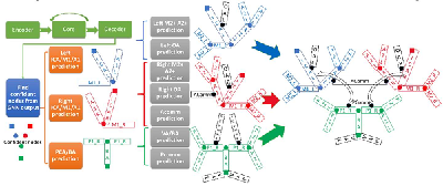
Methods: By semi-automatically tracing arteries based on an open-curve active contour model in a graphical user interface 12 basic morphometric features and 16 basic intensity features for each artery were identified. Arteries were then classified as one of 24 types using prediction from a probability model. Based on the anatomical structures, features were integrated within 34 vascular groups for regional features of vascular trees. Eight 3D MRA acquisitions with intracranial atherosclerosis were assessed to validate this technique.
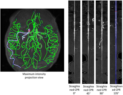
Results: Arterial tracings were validated by an experienced neuroradiologist who checked agreement at bifurcation and stenosis locations. This technique achieved 94% sensitivity and 85% positive predictive values (PPV) for bifurcations and 85% sensitivity and PPV for stenosis, respectively. Up to 1456 features such as length, volume, and averaged signal intensity for each artery, as well as vascular group in each of the MRA images, could be extracted to comprehensively reflect characteristics, distribution and connectivity of arteries. Length for the M1 segment of the middle cerebral artery extracted by this technique was compared with reviewer measured results, and the intra-class correlation coefficient was 0.97.
Conclusion: A semi-automated quantitative method to trace, label and measure intracranial arteries from 3D-MRA was developed and validated. This technique can be used to facilitate quantitative intracranial vascular research, such as studying cerebrovascular adaptation to aging and disease conditions.
Key words: intracranial artery; vessel tracing; feature extraction; Open-Curve Active Contour; magnetic resonance angiogram; time-of-flight
Published, Magnetic Resonance in Medicine (IF: 3.924)
http://onlinelibrary.wiley.com/doi/10.1002/mrm.26961/abstract
DOI: 10.1002/mrm.26961
Youtube channel for demos
https://www.youtube.com/playlist?list=PLxG3HeAddxYb_KQbL2fKNV5BW99yMCksY
Selected for Editor's picks of the journal in June 2018
video for introduction: https://www.
Q&A: https://blog.ismrm.org/2018/06/29/qa-with-li-chen-and-chun-yuan/

admin1
memeda
ssy
 Login
Login